A 1905 Paper Set a Floor Nothing Can See Below
Albert Einstein’s paper “On a Heuristic Point of View Concerning the Production and Transformation of Light” reached the editors of Annalen der Physik on 18 March 1905 and was published that June, one of four papers from what is now called his annus mirabilis [1]. Its actual claim is narrower than the “light is made of particles” shorthand it gets today. Einstein proposed, as a heuristic — his own word, chosen because he was not yet willing to overturn the wave theory that already explained diffraction and interference — that “the energy of a light wave emitted from a point source is not spread continuously over ever larger volumes, but consists of a finite number of energy quanta that are spatially localized at points of space, move without dividing and are absorbed or generated only as a whole” [1]. In his own notation, the energy of one such quantum was the constant ratio Rβ⁄N — built from the gas constant, a term from Wien’s radiation law, and Avogadro’s number — multiplied by the frequency ν. That ratio is the quantity every physics student now writes as Planck’s constant, so the relation reads:
energy proportional to frequency, with nothing smaller possible in between. That single relation is what let Einstein correctly derive the photoelectric effect’s otherwise strange behavior: raising a light’s intensity changes only how many electrons a surface ejects, never their individual energy, while raising its frequency raises that energy directly. It is also, specifically, the achievement the Swedish Academy cited when the 1921 physics prize — reserved a year and awarded in 1922 — went to Einstein “for his services to Theoretical Physics, and especially for his discovery of the law of the photoelectric effect” [2]. Not for relativity, which the committee still treated as unconfirmed. Einstein’s Nobel is for the light quantum, full stop.
That quantum is a hard floor. A detector cannot register three-quarters of a photon at a given frequency; it registers one, or it registers none. Four hundred million years before anyone wrote the equation down, a lineage of light-sensitive cells had already been searched, by mutation and selection, into a device that sits within a small factor of exactly that floor.
Human Rod Vision Needs Only a Handful of Quanta
Selig Hecht, Simon Shlaer, and Maurice Pirenne set out in 1942 to measure that floor directly, publishing in the Journal of General Physiology under conditions built to strip out every avoidable source of loss [3]. Subjects dark-adapted for 40 minutes, then viewed brief flashes — 1 millisecond long, 510 nanometers, chosen to sit at rhodopsin’s absorption peak — aimed at a patch of retina roughly 20 degrees from the fovea, where rods are dense and cones are scarce, through a test spot smaller than 10 arcminutes across [3]. They defined threshold operationally: the energy a subject reported seeing on 60 percent of trials. That threshold worked out to between 54 and 148 quanta of light striking the cornea, depending on the subject [3].
Most of those quanta never reach a rod. Correcting for reflection off the cornea (about 4 percent lost), absorption in the lens and other ocular media (roughly half), and the fraction of transmitted light a rod’s outer segment fails to catch, Hecht and colleagues brought the corneal count down to an estimate of about 5 to 8 quanta actually absorbed by rhodopsin for a reliably detectable flash [3] — a figure later work still cites, essentially unrevised, as roughly 5 to 7 [8]. Because the test spot illuminated several hundred rods at once, each of those absorptions almost certainly landed in a different cell: the visual system was being asked to notice somewhere between five and eight individual rods, each catching exactly one photon, inside a single flash. That is not “detecting light” in any loose sense. That is counting quanta.
A Suction Electrode Showed the Single Photon Directly
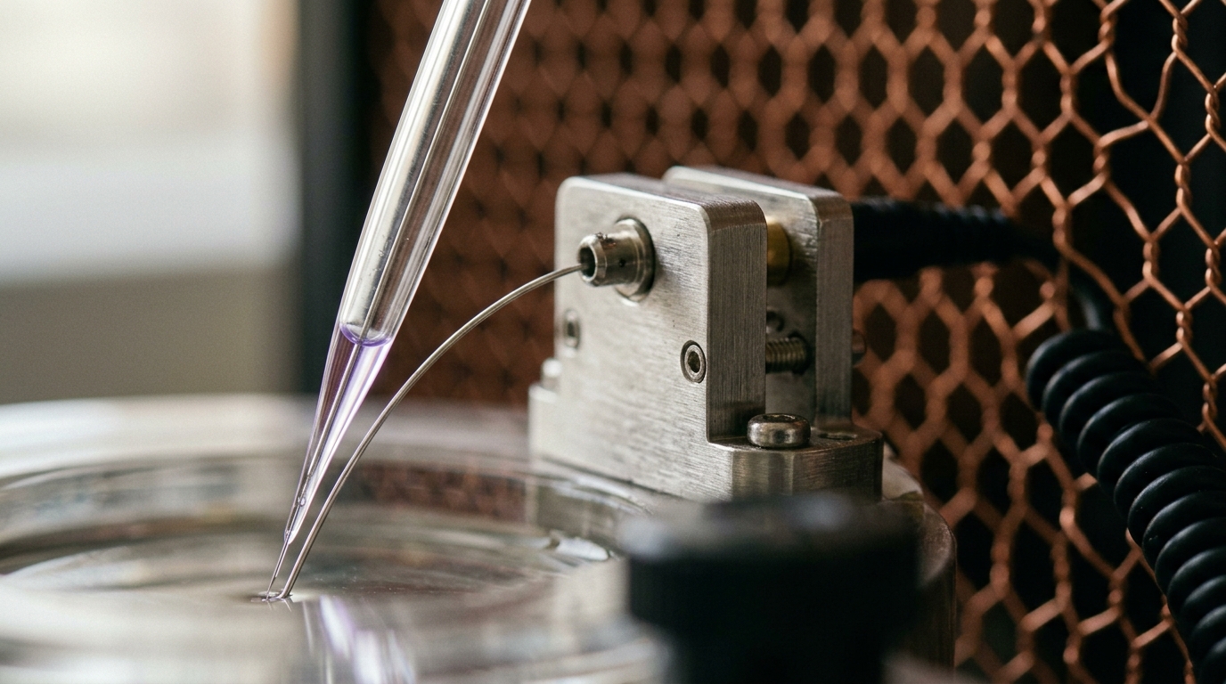
Hecht’s number was inferred from what people reported seeing; it said nothing about what one rod actually did with one photon, and it could not by itself rule out some population-level averaging trick standing in for true single-cell sensitivity. Denis Baylor, Trevor Lamb, and King-Wai Yau closed that gap in 1979, recording in the Journal of Physiology from single toad rod outer segments drawn into the tip of a fine glass suction electrode, a method that measures the current across one cell’s membrane directly [4]. Under dim, calibrated flashes, the current traces did not vary continuously with flash strength the way a graded chemical response would. They clustered: amplitude histograms showed a peak at zero and a second peak near one picoampere, and the fraction of trials landing in that second peak rose and fell with flash intensity exactly as expected if each discrete bump corresponded to a single photon isomerizing a single rhodopsin molecule [4]. The single-photon response was no longer a statistical inference. It was a trace on an oscilloscope.
What makes that trace visible at all is amplification most catalysts never manage. A photoisomerized rhodopsin molecule activates a run of copies of the G-protein transducin; each activated transducin turns on a phosphodiesterase molecule that hydrolyzes cyclic GMP near the diffusion limit; falling cyclic GMP closes many membrane channels at once. Marie Burns and Denis Baylor’s 2001 review describes the net result — one photoisomerization interrupting the flow of on the order of 10^5 or more sodium ions into the cell — as “particle amplification” built from exactly those three multiplying steps [5]. The middle step’s precise size is still disputed: light-scattering measurements from the 1980s put the rate of transducin activation at 700 to 1,000 per second, while later biochemical re-measurements, correcting for how badly earlier preparations disrupted the rod’s internal membrane structure, put it closer to 120 to 150 per second — a discrepancy Burns and Baylor call, in their own words, still “puzzling” two decades on [5]. Not in dispute is the overall multiplication: independent estimates converge on a combined gain of roughly 10^5 to 10^6 across rhodopsin and phosphodiesterase together, enough that one absorbed photon produces a current change an electrode reads cleanly against baseline noise [6].
More striking than the size of the amplifier is its precision. A cascade this large, triggered by one molecule’s random encounter with one photon, has every reason to be noisy — each downstream biochemical step is itself a random process. Yet single-photon response amplitudes recorded from the same rod are remarkably uniform trial to trial. Rieke and Baylor’s separate 1998 study in Biophysical Journal tested the obvious explanations for that uniformity — saturation somewhere in the cascade, calcium feedback — and ruled both out; the reproducibility, they concluded, more likely comes from rhodopsin’s own catalytic activity shutting off through a sequence of several distinct steps rather than one, so the molecule’s total active lifetime is the sum of several random intervals rather than a single one, which narrows around its average the way any such sum does [7]. A single molecule’s activity has effectively been shaped, by selection rather than design, to behave almost as reliably as an average over many.
Humans Can Report a Single Photon, but Barely
Baylor’s electrode measured a rod; it did not ask whether a person, consciously, could notice one photon rather than five. That is a harder experiment: it needs a light source that reliably delivers exactly one photon and a psychophysical protocol sensitive enough to find a signal that faint. Jonathan Tinsley and colleagues built both in 2016, combining a quantum light source with a two-alternative forced-choice task: three trained observers judged which of two one-millisecond intervals had contained a photon, then rated their confidence, across 30,767 trials, of which 2,420 were true single-photon presentations [8]. Averaged across every trial, the probability of a correct response was 0.516 ± 0.010 — barely above the chance level of 0.5, and, at P = 0.0545, not quite past the conventional threshold for statistical significance on its own [8]. Restricting the analysis to trials where subjects reported high confidence sharpened the result considerably: correct-response probability rose to 0.60 ± 0.03, P = 0.0010 [8]. The honest reading is not that people reliably see single photons in any everyday sense — they do not — but that the visual system carries a detectable, reproducible trace of one quantum event all the way to a forced decision, some of the time, which is itself remarkable given how many stages of noisy amplification and neural pooling that trace has to survive.
Part of that difficulty is a floor the rod imposes on itself, deliberately. Rhodopsin’s chromophore, 11-cis-retinal, occasionally isomerizes with no photon involved, and this thermal event triggers the identical amplification cascade a real photon would — a false alarm indistinguishable from a true signal at the point where the cell reports it. Burns and Baylor cite a rate constant for this in toad rods, near 20°C, of about 10^-11 per second per rhodopsin molecule, “corresponding to an average wait to isomerization of 2000 years” for any single molecule [5]. Baylor, Nunn, and Schnapf measured the population-level consequence directly in primate rods: discrete dark events resembling single-photon responses occur at roughly 0.006 per second per rod, on the order of one spurious event every few minutes in a cell that is otherwise silent [9]. That rate is tolerable only because it is so low relative to how rarely a dim scene delivers a genuine photon to any one rod; raise it even modestly and false alarms would swamp real detections. It is also why the visual system never trusts a single rod’s single bump as the basis for a percept: Hecht’s five-to-eight-quantum threshold and Tinsley’s confidence-gated result are both, functionally, downstream decision rules that require several independently generated events to agree within a short window before a signal counts as real — the criterion a rational detector should adopt when a true event and a false one look identical at the source and differ only in how often each occurs [6]. Reliability at this limit is bought with a coincidence requirement, not with an ever-lower threshold.
Selection Repeated the Trick From Insects to the Deep Sea
Rod vision at the quantum floor is not a vertebrate curiosity; it recurs wherever a lineage’s survival has depended on seeing in near-darkness, arrived at independently each time. Eric Warrant’s 2017 review in Philosophical Transactions of the Royal Society B catalogs several cases: the nocturnal sweat bee Megalopta genalis navigates rainforest understorey at night while absorbing fewer than five photons per photoreceptor per second, far below what its diurnal relatives could use [10]. The cockroach Periplaneta americana can detect a moving object while absorbing fewer than one photon per photoreceptor every 10 seconds [10]. The elephant hawkmoth Deilephila elpenor keeps motion vision functional down to overcast-starlight intensities by pooling: Warrant’s review estimates each visual channel spatially summing signals from roughly 109 neighboring ommatidia, a strategy extending usable sensitivity on the order of a hundredfold at a direct cost to spatial resolution [10].
The deep sea runs the identical trade inside a vertebrate eye. Fanny de Busserolles and Justin Marshall’s 2017 review of lanternfish retinas reports the highest rod density recorded for any vertebrate, packed into photoreceptors of the smallest diameter known in either vertebrates or invertebrates, feeding a rod-to-ganglion-cell convergence ratio of 200 to 600 to 1 — against roughly 1 to 2 in the human fovea [11]. That pooling buys lanternfish an estimated 10 to 100 times the optical sensitivity of a human eye to a dim, extended source, at a cost the review states without softening: spatial resolving power of only 1.6 to 4.8 cycles per degree, among the lowest recorded for any deep-sea fish [11].
| Lineage | Photon regime tolerated | Strategy | Price paid |
|---|---|---|---|
| Human rod, dark-adapted | ~5–8 quanta absorbed for reliable detection [3] | Single-photon-sensitive rod; downstream coincidence pooling | Rod signal is slow and achromatic |
| Nocturnal sweat bee | <5 photons per photoreceptor per second in flight [10] | Wide-aperture ommatidia, neural summation | Reduced spatial resolution |
| Nocturnal cockroach | <1 photon per photoreceptor per 10 s [10] | Extreme temporal summation | Very slow motion detection |
| Deep-sea lanternfish | 10–100× human optical sensitivity to extended sources [11] | Peak rod density; 200–600:1 rod-to-ganglion pooling | Acuity of only 1.6–4.8 cycles/degree |
Three lineages — an insect compound eye, a deep-sea vertebrate eye, a primate eye — arrived by entirely separate evolutionary routes at the same short list of tricks: enlarge the collecting aperture, extend the integration time, pool signals across many detectors. None found a way around the photon. Each found its own way to live as close to it as the physics of shot noise allows.
Selection Found a Physical Constant and Stopped
Every piece of this record points the same direction from a different angle. Einstein’s 1905 paper defined the smallest parcel light can be split into [1]. Hecht, Shlaer, and Pirenne showed a dark-adapted human eye needs only a handful of those parcels to register a flash [3]. Baylor, Lamb, and Yau showed a single rod resolving exactly one, run through a biochemical amplifier precise enough that its output barely varies from trial to trial [4, 7]. Tinsley and colleagues pushed the same chain all the way to a conscious forced choice and found a real, if statistically fragile, signal at the single-photon limit itself [8]. And at every step the honest limits — thermal noise indistinguishable from a genuine photon, a coincidence requirement standing in for perfect sensitivity, acuity traded away for gain in every lineage that has tried — describe a system straining against a wall it cannot get past, not one that never needed to.
That wall was not put there by biology. It is the shot noise built into the discreteness of light itself, the same discreteness Einstein wrote down as a heuristic in 1905 and that the 1921 Nobel committee credited without yet believing everything else he had proposed [2]. Natural selection did not derive that constant, has no mechanism for representing it symbolically, and could not have aimed at it on purpose. What four hundred million years of retinal variation and culling did was search a space of possible photoreceptors long enough, and hard enough, to land within a small factor of a number selection could never have written down — and then stop, because past that number there was nothing left to find.