A Pathologist Removed the Brain Before Permission Existed to Give
Albert Einstein died shortly after 1 a.m. on 18 April 1955, at Princeton Hospital, of a ruptured abdominal aortic aneurysm, at the age of seventy-six [3]. Within hours the examining pathologist, Thomas Harvey, removed his brain, weighed it at 1,230 grams, took forty measurements, and perfused it with formalin [3]. The archival finding aid kept by the National Museum of Health and Medicine states plainly what the tidier version of the story usually skips: “No permission for the removal and preservation had been given by Einstein or his family, but when the family learned about the study, permission to proceed with the study was granted as long as the results were only published in scientific journals and not sensationalized” [7].
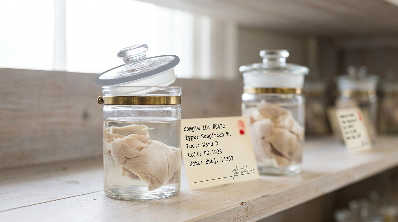
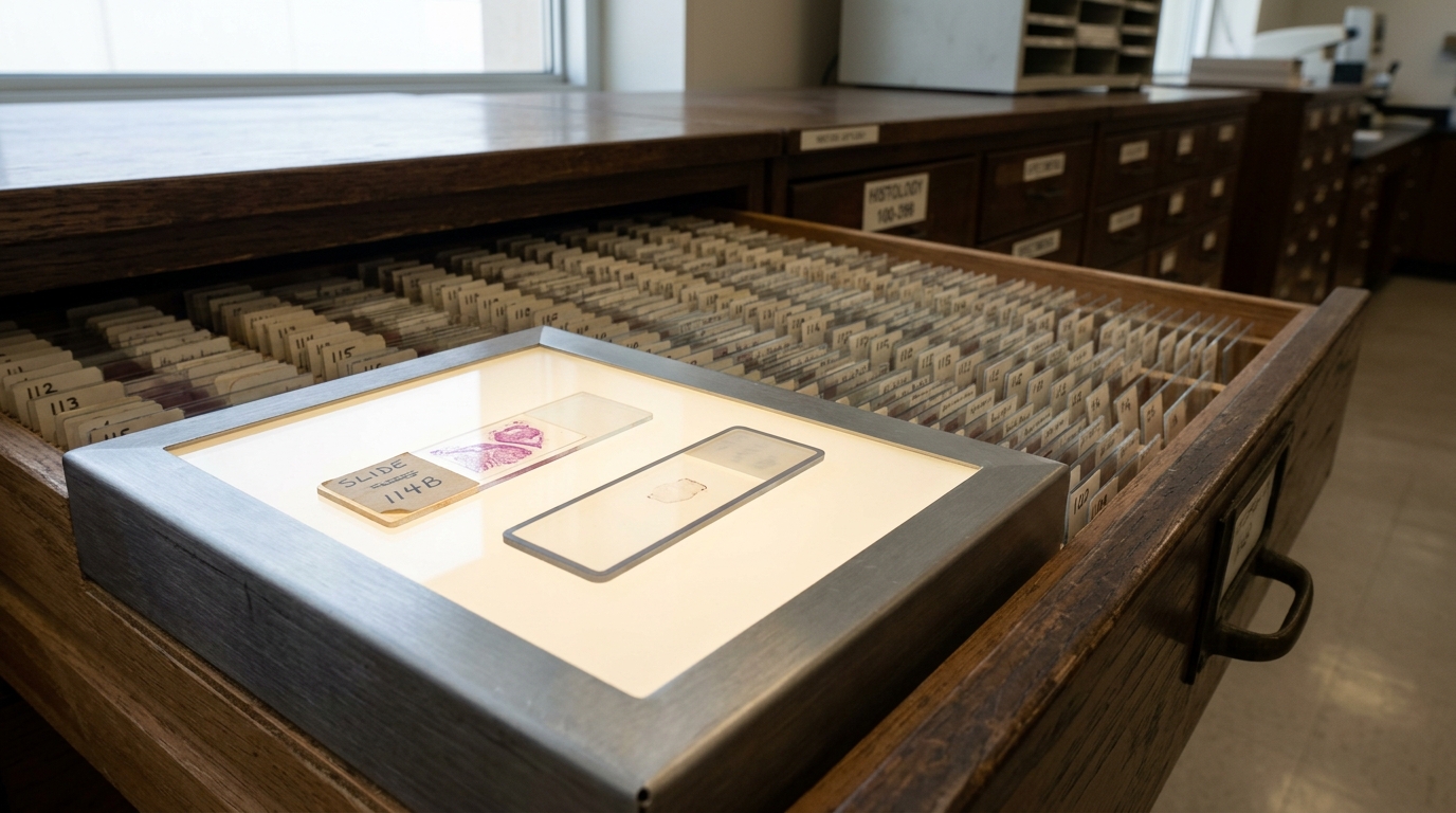
Harvey had the cerebral hemispheres cut into roughly 240 blocks of about ten cubic centimetres each, embedded them in celloidin, and recorded each block’s original location on a hand-drawn “roadmap” [1]. He kept two full sets of the resulting slides for himself and distributed the rest to what the museum’s own records call “handpicked leading pathologists of the time” [7]. Over the following decades, no fewer than eighteen investigators received tissue or photographs from him directly [3]. The brain itself mostly did not travel to a lab; it traveled with Harvey — away from Princeton and eventually to a medical testing lab in Wichita, Kansas, where in August 1978 New Jersey Monthly reporter Steven Levy tracked him down and published “I Found Einstein’s Brain,” reviving a story most people had assumed was long finished [7].
By the time Falk, Lepore and Noe went looking for the original whole-brain photographs in 2010, the trail had gone cold the way an uncatalogued relic’s trail goes cold: the largest surviving set of celloidin blocks, 180 of the original 240, sat at Princeton’s University Medical Center; the largest surviving set of slides, 567 of them, sat at the National Museum of Health and Medicine; and smaller fragments turned up, by Falk’s own accounting, in Ontario, California, Alabama, Argentina, Japan, Hawaii, and Philadelphia [3]. A 1988 attempt to sequence DNA from one celloidin block found it “completely fragmented, completely denatured” [3] — the tissue degrading even as claims made about it multiplied.
None of this makes Harvey a simple villain: contemporary biographies record that Einstein had “insisted that his brain should be used for research” [1], and that he had already, in 1951, joined other physicists in undergoing electroencephalographic recordings purely for research purposes [1]. Einstein was, on the available evidence, genuinely sympathetic to being studied. But a 2018 historiographical review of the collection notes that Hans Albert Einstein’s actual condition was narrower than that blanket endorsement suggests — permission granted only if results appeared in “scientific journals of high standing” [8] — and that Harvey was later “severely criticized” over a “sensationalized account” of having, literally, taken parts of the brain “on the road” [8]. Wanting one’s brain studied, consenting to EEG recordings while alive, and consenting to four decades of a private pathologist’s personal custody, a cross-country relocation in mason jars, and informal distribution to whoever wrote a persuasive letter are different things. The record supports only the narrower version.
Four Studies Examined the Same Tissue and Reached Incompatible Conclusions
Six peer-reviewed papers eventually came out of Harvey’s collection [3]. Four carry the weight of the popular “Einstein’s brain was special” story, and set side by side, they do not agree with each other.
Diamond and colleagues compared four cortical regions — areas 9 and 39, both hemispheres — from Einstein’s brain against the same regions in eleven male control brains, whose donors had died at an average age of sixty-four, twelve years younger than Einstein, and reported one statistically significant result: a lower neuron-to-glia ratio in left area 39, meaning more glial cells per neuron than the controls showed [2]. Witelson, Kigar and Harvey compared Einstein’s whole, unsectioned brain against thirty-five male brains held in the Witelson Normal Brain Collection (mean IQ 116) and reported that his brain weighed 1,230 grams against a control mean of 1,400 grams — lighter, not heavier — but that each parietal region measured fifteen per cent wider than the control average, and that the posterior ascending branch of the Sylvian fissure ran directly into the postcentral sulcus in both hemispheres, which they read as meaning no parietal operculum was present at all [1]. Their threshold for “statistically significant” was any measure lying at least two standard deviations from the control mean [1], applied without any stated correction across roughly two dozen separate measurements listed in their own results table [1].
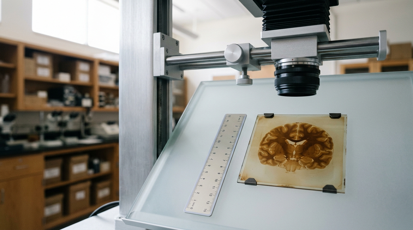
Fourteen years later, working from photographs of the same brain rather than the by-then-dispersed tissue itself, Falk, Lepore and Noe directly contradicted Witelson’s signature claim: “Einstein’s brain is not spherical, does not lack parietal opercula and has non-confluent Sylvian and inferior postcentral sulci” [3]. Their own positive findings were different again — an “extraordinary prefrontal cortex,” a face-and-tongue motor region “greatly expanded” in the left hemisphere, and a fourfold transverse occipital sulcus pattern they call “extremely rare, if not unique” [3] — reached by comparing Einstein’s cortex, qualitatively, against sulcal descriptions from two older published atlases of sixty and twenty-five human brains, not a purpose-built control sample matched to Einstein’s age or sex [3].
Terence Hines’s 2014 review stepped back from any single paper to ask whether the underlying pattern held up. On Diamond’s finding specifically, he noted that the study amounted to twenty-eight separate comparisons — seven measures across four regions — of which exactly one crossed the conventional threshold, calling that result “not surprising” under ordinary chance alone [4]. Reviewing the histological literature as a whole, his conclusion was blunt: those studies “have, in spite of claims to the contrary, found essentially no differences between his brain and that of controls” [4]. His proposed remedy was the one none of the earlier teams had used: blind the comparison, so a researcher scoring slides or photographs does not already know which specimen is supposed to be extraordinary [4].
| Study | What was compared | Central claim | Documented problem |
|---|---|---|---|
| Diamond et al., 1985 | 4 regions, Einstein vs. 11 controls (avg. 12 yr younger) | Higher glia:neuron ratio, left area 39 | 1 of 28 comparisons significant; full cell counts never published |
| Witelson et al., 1999 | Whole brain, Einstein vs. 35 controls | Missing parietal operculum; parietal lobes 15% wider | Significance set at 2 SD, uncorrected, across ~24 measures |
| Falk et al., 2013 | 14 photographs vs. two published atlases | Operculum present; expanded prefrontal and motor cortex | Qualitative reading against unmatched historical samples |
| Hines, 2014 | Re-review of the three above | Findings do not replicate; consistent with chance | Recommended blinding never adopted by the original teams |
A Sample Size of One Cannot Carry the Weight These Claims Put on It
Every one of these papers compares a control group of a few dozen brains against exactly one brain belonging to a famous man, and that asymmetry is the whole problem, not a detail of it. A control group can establish a range of ordinary variation; it cannot, on its own, tell you whether one additional case falling near the edge of that range is genuinely unusual or simply where, given wide enough natural variation, someone eventually was going to land. Witelson’s own paper reports that Einstein’s brain showed “moderate atrophy… around the main fissures in the central regions in both hemispheres, to an extent common for a person in their eighth decade” [1] — a reminder that the tissue being read for genius was also, unremarkably, an old man’s brain.
None of the four studies pre-registered which measurement would count as evidence of exceptional intellect before looking at the data. Witelson’s team came closest to a stated hypothesis, reasoning from Einstein’s own account of his thinking as “associative play” of “more or less clear images” of a “visual and muscular type” [1] — a genuinely reasonable starting point for a study design. But a hypothesis phrased that broadly can be confirmed by almost any subsequent anomaly in a large, functionally diffuse brain region: a missing structure, an enlarged one, an unusual fold would each have counted as support. That elasticity, combined with base-rate neglect — the plain fact that any two brains, compared across dozens of measurements, will differ substantially somewhere by chance alone, because normal anatomical variation between individual human brains is large — is what let four independent research teams each find a different “special” feature in the same tissue, and none of them find the same one twice.
The comparison groups compound the problem rather than fixing it. Diamond’s eleven controls, Witelson’s thirty-five, and Falk’s borrowed atlases of sixty and twenty-five brains were never assembled to test a shared, pre-specified hypothesis about Einstein specifically; each was whatever normal-brain collection happened to be on hand when a given team went looking. A control group built after the fact, from whatever specimens are available, cannot police multiple comparisons the way a control group designed in advance for one stated question can — it answers “was Einstein’s brain, in some sense, at the edge of a distribution?” while quietly leaving unaddressed which edge, of how many measured distributions, should have counted in advance.
Evolutionary Neuroscience Finds No Organ Built for Genius
The alternative to relic-hunting is asking what evolutionary and developmental neuroscience actually predicts about where intelligence lives in a brain, and the honest answer does not point toward any single traceable structure. Pietschnig and colleagues’ 2015 meta-analysis, pooling 148 samples and more than 8,000 people across 88 studies, put the correlation between brain volume and IQ at r = 0.24 — roughly six per cent of shared variance — and concluded that brain volume “plays only a minor role in explaining IQ test performance in humans,” adding that even this modest figure is likely inflated by publication bias in the earlier literature [5]. Witelson’s own numbers make the same point from the opposite direction: at 1,230 grams, Einstein’s brain sat below, not above, the 1,400-gram control mean, and her paper states plainly that this result was “clearly indicating that a large (heavy) brain is not a necessary condition for exceptional intellect” [1].
Nor does modern behavior genetics locate intelligence in one part or one gene. Plomin and von Stumm’s 2018 review describes genome-wide association studies that have identified variants jointly accounting for roughly twenty percentage points of intelligence’s approximately fifty per cent heritability, aggregated across thousands of individual DNA variants, each of tiny effect [9]. A trait built from thousands of small, distributed genetic contributions acting through diffuse developmental pathways is not a trait with one anatomical seat waiting in a jar of formalin; structurally, it is closer to the opposite kind of trait.
What evolutionary neuroscience does robustly support is plasticity, not innate anatomical destiny. Maguire and colleagues scanned sixteen licensed London taxi drivers — mean 14.3 years’ experience navigating the city’s street network from memory, a skill acquired over roughly two years of training known as “the Knowledge” — against fifty non-driving controls, and found significantly larger posterior hippocampal gray matter in the drivers, positively correlated with years spent driving, alongside a comparatively larger anterior hippocampal region among the controls [6]. That is a real, structural change driven by sustained use of an entirely ordinary brain region, not the discovery of a rare structure that predicted navigational talent in advance. Applied back to Einstein, the same logic cuts against every one of the genius-anatomy papers: even a real, replicated anomaly in his cortex would leave open whether decades spent doing a specific kind of visuospatial work reshaped ordinary tissue, rather than unusual tissue having enabled the work — a direction-of-causation question no single postmortem brain, examined once, at seventy-six, could ever settle.
The Episode Was Relic-Worship Wearing a Lab Coat
Strip away the vocabulary of neuroscience and the shape of the story is an old one: a body part taken from a revered figure without full authorization, divided into venerated fragments, circulated among a scattered priesthood of specialists, carried across the country by its keeper for decades, and periodically rediscovered to renewed public fascination — Levy’s 1978 exposé functioning, structurally, exactly like the rediscovery of a saint’s relic in a forgotten reliquary. A secular, scientifically literate culture did not abandon that instinct; it built the reliquary out of celloidin blocks and peer-reviewed journals instead of gold and stained glass.
What makes this literature useful, in the end, is not any of its four incompatible positive findings — it is Hines’s negative one. A relic has to be found special every time someone looks, because that is the entire premise of keeping it; a well-run study has to be willing, most of the time, to find nothing, and to say so when it does. Blinding the comparison, pre-registering the measurement, gathering more than one case before generalizing about “genius” — every fix Hines names is a way of making it possible for the answer to come back negative. That the record from Harvey’s autopsy table produced four different positive stories and one careful reviewer concluding that none of them survive scrutiny [4] is not a failure of the search. It is what an honest search looks like when the thing being searched for — one organ, in one man, that made him a genius — was never there to find.