Forty to sixty-five separate inventions was the reasonable reading of the anatomy
When Leon von Salvini-Plawen and Ernst Mayr surveyed the animal kingdom’s light-sensing organs in 1977, they were not being careless. Their published conclusion was that photoreceptors of various degrees of differentiation had evolved independently “in at least 40, if not 65 or even more separate phyletic lines” [1]. That figure gets repeated today as a curiosity, a large round number meant to sound impressive. It was not a round number and it was not a guess. It came from two independent lines of comparative evidence that, in 1977, pointed the same way and reinforced each other.
The first line was optical. A vertebrate or cephalopod camera eye focuses light through a single refracting lens onto a continuous retina, the way a photographic camera does. An arthropod compound eye instead tiles hundreds to thousands of separate optical units, each an independent lens-and-photoreceptor bundle sampling one narrow direction, so spatial resolution comes from combining many partial views rather than from one continuous image. A scallop’s eye is neither of those: Pecten builds a concave mirror behind its retina and forms an image by reflection, the same principle later used in reflecting telescopes. These are not minor variations on a single plan. They are three different solutions to the problem of forming an image from light, built by lineages that comparative anatomy had already placed on separate branches long before any of them possessed eyes.
The second line compounded the first: embryonic origin. The vertebrate eye develops as an outgrowth of the neural tube itself, so the retina is, developmentally, brain tissue, wired in an inverted arrangement in which photoreceptors face away from incoming light, behind a layer of nerve fibres the light must cross first. The cephalopod eye, superficially so close in its camera layout that Darwin used the comparison directly, develops instead from surface ectoderm, in the everted arrangement most other invertebrate eyes share, photoreceptors facing straight toward the light. Two organs that converge almost perfectly on one optical solution turn out to be assembled from different starting tissue, wired in mirror-image orientation, by lineages with no eye-bearing common ancestor. Extend that logic across every eye-bearing phylum, and stack the count of genuinely distinct optical principles on top of it, and Salvini-Plawen and Mayr’s range falls out: not fewer than forty separate origins, plausibly sixty-five or more.
For its time this was close to a working consensus, and it did real explanatory work. It explained why comparative anatomists could never draw one continuous lineage tree for eye designs the way they could for, say, tetrapod limbs. It explained why molecular biologists going into the 1980s had no particular reason to expect that a fly’s compound eye and a mammal’s camera eye would share underlying genetic machinery — nothing in the anatomy or the embryology predicted it. What follows is the specific pair of experiments that overturned that expectation, and it matters to be precise about which one did what, because the popular retelling compresses two distinct papers, a year apart, into a single moment of discovery it was not.
One paper found the shared gene; a second, a year later, proved what it could do
The Drosophila mutation eyeless had been known since 1915, when it was first described from its phenotype: flies homozygous for hypomorphic eyeless alleles have reduced or absent compound eyes, though their simple eyes, the ocelli, are unaffected [3]. For nearly eighty years that was all it was — a named phenotype with no known molecular identity. In 1994, Rebecca Quiring, Uwe Walldorf, Urs Kloter and Walter Gehring cloned the gene. It encodes a transcription factor carrying both a paired domain and a homeodomain, and its DNA sequence showed extensive homology to the mouse Pax-6 gene, known from the Small eye mutant, and to the human Aniridia gene [2]. Two independently identified transposable-element insertions, in the alleles ey² and eyR, mapped into this cloned sequence and disrupted its expression specifically in the eye primordia, confirming that the gene really was eyeless [2]. The paper’s own summary of the implication was direct: eye morphogenesis appeared to be under similar genetic control in vertebrates and insects, “in spite of the large differences in eye morphology and mode of development” that had grounded Salvini-Plawen and Mayr’s count in the first place [2].
Sequence homology alone invites an obvious objection: genes get reused for unrelated jobs across evolution constantly, and a shared ancestral protein does not by itself prove a shared ancestral function. Homology of the gene is not the same claim as homology of the organ, and by 1994 nobody had shown that the fly and mouse copies of this gene actually did the same thing when it mattered. That gap is what Georg Halder, Patrick Callaerts and Walter Gehring closed in 1995, in the paper that is usually, and imprecisely, credited with the whole discovery.
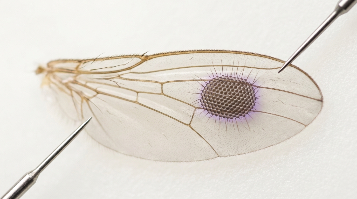
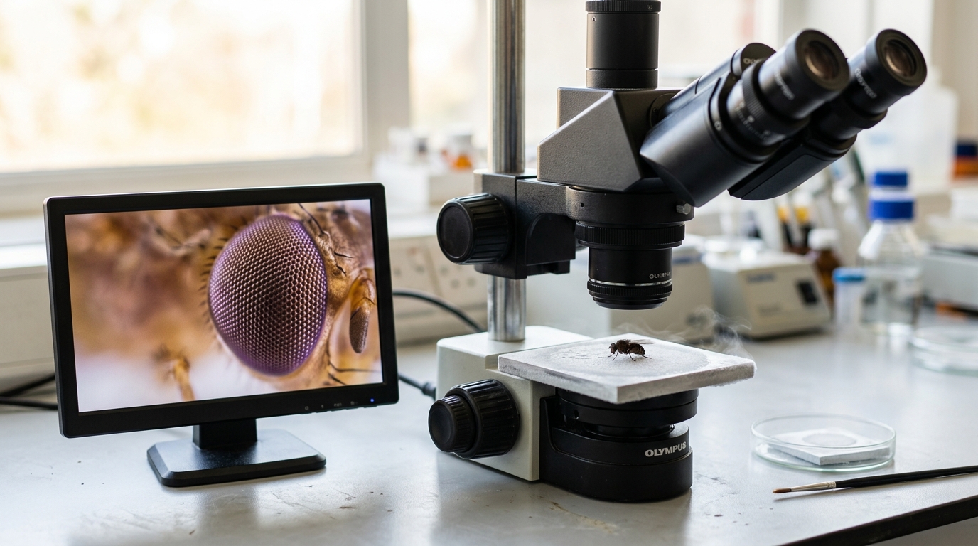
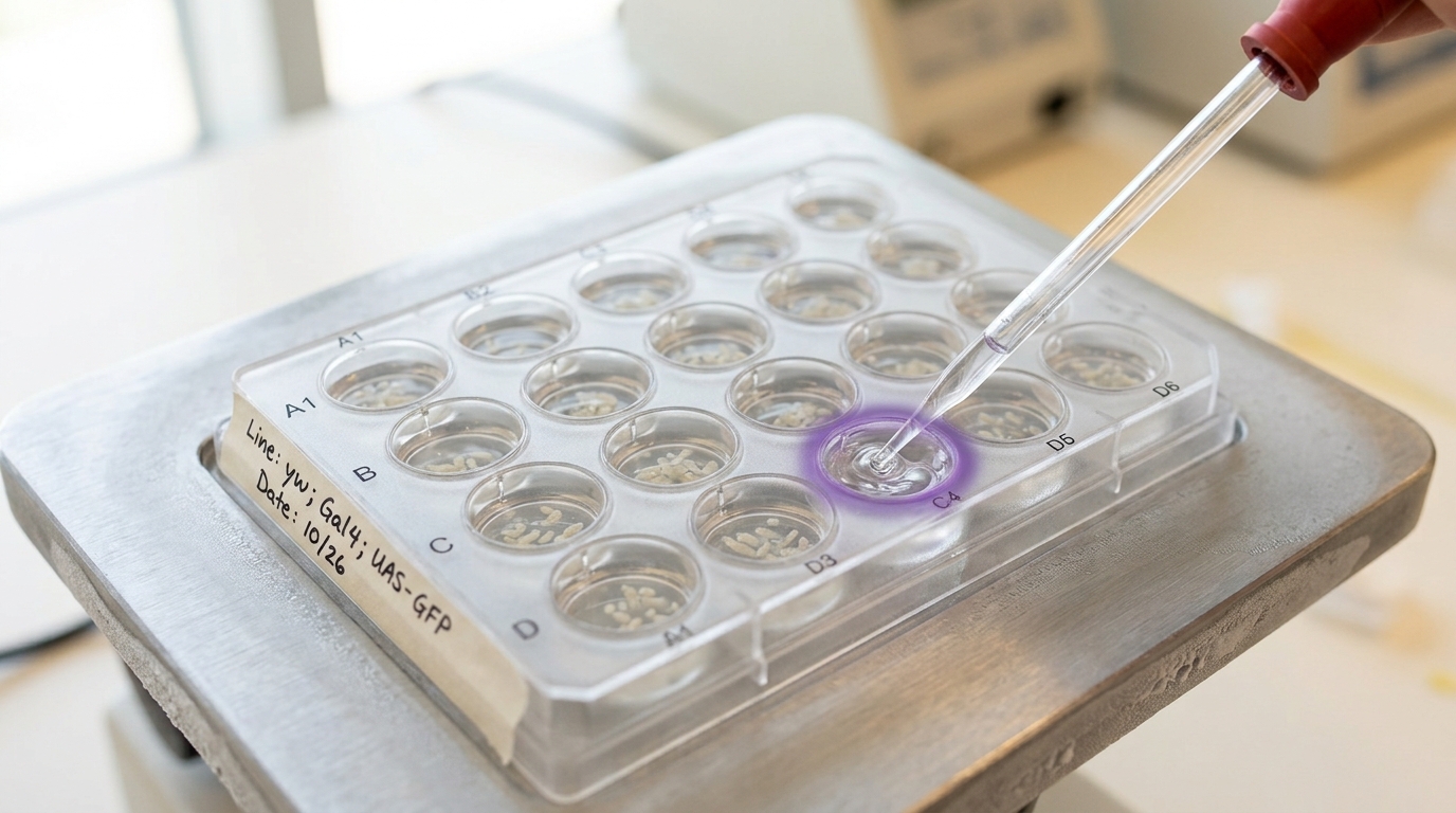
Halder, Callaerts and Gehring used the GAL4-UAS system — a yeast transcriptional activator crossed into a target line carrying the eyeless coding sequence behind a GAL4-responsive promoter — to force eyeless expression in imaginal disc tissue where it is never normally switched on: the wing, leg and antennal discs [3]. Of roughly twenty GAL4 driver lines they tried, only three gave viable adult flies to examine in detail, a detail worth holding onto for what it implies about “sufficiency,” which the next section returns to [3]. In the flies that did survive, ectopic eye structures formed on the wings, on all six legs, on the antennae and on the halteres, fully penetrant in the surviving genotypes [3]. These were not blemishes or disorganised tissue. Scanning electron microscopy showed hexagonal ommatidia and interommatidial bristles organised almost exactly as in a normal eye; histological sections found a complete cornea, cone cells, and primary, secondary and tertiary pigment cells; antibody staining against the neuronal marker ELAV showed photoreceptors differentiating in the correct sequence, with a trapezoidal array of rhabdomeres matching a normal ommatidium [3]. A wing-borne ectopic eye typically carried around 350 facets, against roughly 800 in a normal compound eye — smaller, but built from the same parts in the same arrangement [3].

The result that makes this a genuinely cross-phylum experiment, rather than a fly gene doing a fly-specific trick, sits in the same paper, not a separate one. Halder, Callaerts and Gehring also cloned the full-length mouse Pax-6 cDNA and drove it with the identical GAL4 line, in Drosophila, in place of the fly’s own eyeless sequence [3]. The mouse gene induced ectopic eye structures too, with ommatidial arrays and interommatidial bristles closely resembling those produced by the fly gene [3]. The paper is explicit about the detail that gives this its interpretive force: “the ectopic eye structures thus formed contain Drosophila-type ommatidia and not mouse-type ommatidia” [3]. The mouse protein did not import any vertebrate eye architecture into the fly. It switched on the fly’s own downstream cascade — the network eyeless normally sits atop, including subordinate genes like sine oculis — and that cascade then built the only kind of eye the surrounding fly genome knew how to build. What crossed 500 million years of divergence was the switch, not the blueprint downstream of it. The authors’ own comparison was to Hans Spemann’s classical transplantation experiments, in which grafting the optic-cup primordium to an ectopic site in an amphibian embryo could induce an eye there too [3] — except that this time the inducing signal was a single named gene, and it worked when swapped in from a different phylum entirely.
The two papers, read precisely, establish two different and complementary things. Quiring and colleagues established that the same gene, by sequence, sits behind the eyeless phenotype in flies and the Small eye and Aniridia phenotypes in mice and humans [2]. Halder, Callaerts and Gehring established, a year later, that this shared gene is not just structurally similar but functionally interchangeable at the top of the switch, in both directions of the fly-mouse comparison, and that the resulting organ takes its detailed form from whatever tissue receives the signal [3]. Salvini-Plawen and Mayr’s count of independent eye types was never wrong about the eyes. It was the assumption sitting underneath it — that radically different eyes implied radically unrelated genetic starting points — that the two papers together disproved.
Deep homology names the shared circuitry — Gehring’s strongest claim about it is not the same thing as the concept itself
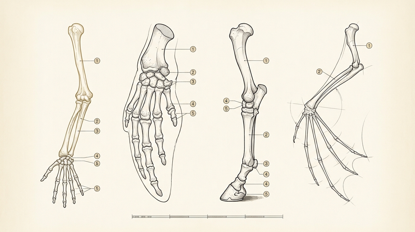
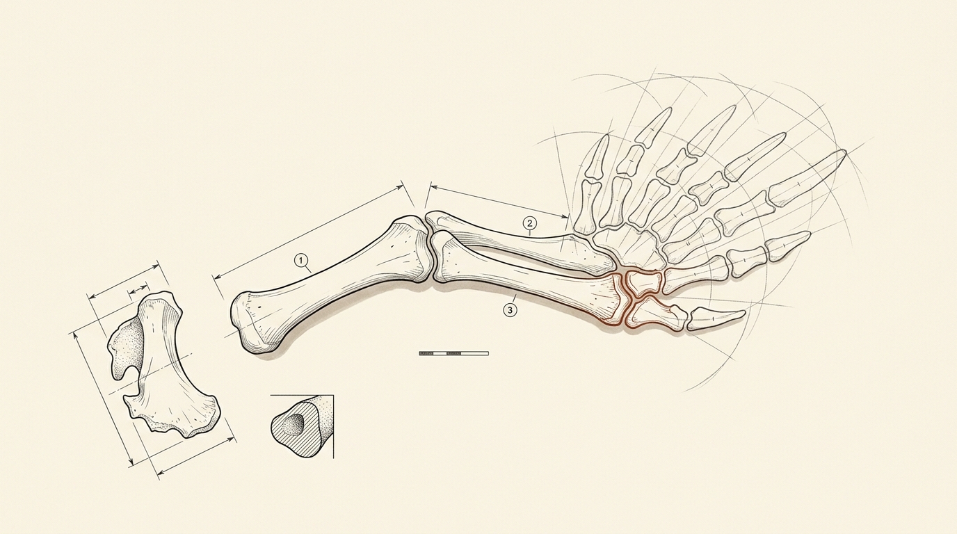
The vocabulary that eventually organised this finding, “deep homology,” was coined by Neil Shubin, Cliff Tabin and Sean Carroll, first in a 1997 review of limb evolution and then in a widely cited 2009 update that folded the eye result into a broader pattern [5, 6]. Their 1997 paper was not about eyes at all. It traced how modifications to a shared set of patterning genes, expressed along the proximodistal axis of a developing appendage, correlate with number and architecture across tetrapod limbs, arthropod legs and insect wings, arguing that a similar underlying regulatory system was established once, in a common ancestor, rather than separately invented by each lineage that grew an appendage [5]. Grace Panganiban and colleagues supplied the specific molecular evidence the following same year: the gene Distal-less, or its vertebrate counterpart Dlx, is expressed along the proximodistal axis of appendages in six different coelomate phyla — polychaete annelid parapodia, onychophoran lobopods, ascidian ampullae and even echinoderm tube feet, alongside arthropod legs and vertebrate limbs — and the pattern of expression across such distant relatives supports a single origin for that regulatory deployment in a pre-Cambrian common ancestor, rather than six separate co-options [13].
By 2009, Shubin, Tabin and Carroll folded eyes, limbs and even beetle horns into one statement of the general principle: “the deep homology of generative processes and cell-type specification mechanisms in animal development has provided the foundation for the independent evolution of a great variety of structures” [6]. Read that sentence slowly, because its two halves are doing different jobs. “Deep homology of generative processes” is the homology claim — the toolkit genes themselves, Pax6 among them, and the cell-type specification programs they sit atop, are inherited from a shared ancestor. “Independent evolution of a great variety of structures” is the convergence claim — the actual organs, compound eyes, camera eyes, tetrapod limbs, insect wings, really were assembled separately, by separate lineages, under separate selective pressures. Deep homology is the claim that both halves are true simultaneously, of the same case, and that is precisely why it is not simply a restatement of either “convergent” or “homologous.”
Gehring’s own reading of this, laid out in a 2005 review, pushes further than Shubin, Tabin and Carroll’s formulation and is worth stating as his claim rather than as settled fact. He argues that because Pax6, together with the six1 and six3 genes, controls eye development across organisms “ranging from planarians to humans,” and because a transcription factor’s conservation across that entire span is not required by function alone — since transcription factors can in principle be swapped in to activate any target gene with the right regulatory elements — the shared control itself is better explained by common ancestry than by convergent re-selection of the same molecule for the same job [7]. His proposal is that a minimal proto-eye, the two-cell-type eye Darwin himself once sketched as a plausible starting point — one photoreceptor cell and one pigment cell — came under Pax6’s control once, in a single bilaterian ancestor, and every subsequent eye type since has been built by progressively intercalating new genes between that original master switch and the structural genes, like rhodopsin, that sit at the bottom of the cascade [7]. He calls this intercalary evolution, and proposes gene duplication and enhancer fusion as the two mechanisms most likely to insert new genes into the growing hierarchy [7]. On this reading, the various eye types are not forty-plus independent inventions in any meaningful sense; they are one invention, elaborated forty-plus different ways.
That is a strong claim, and three separate bodies of evidence complicate it without simply refuting it. First, Pax6 is not an eye-specific gene in the organisms that use it for eyes. Lucie St-Onge and colleagues showed in 1997 that Pax6 knockout mice fail to develop glucagon-producing alpha cells in the pancreas, meaning the same transcription factor is independently essential for endocrine-cell differentiation in an organ with no developmental connection to vision [11]. A gene this pleiotropic is not, by nature, “the eye gene”; its use in eye development is one deployment among several, which sits uneasily with language implying the gene exists to build eyes. Second, sufficiency was never unconditional, even in the paper that demonstrated it: seventeen of the roughly twenty driver lines Halder, Callaerts and Gehring tried did not yield viable, examinable ectopic eyes at all [3], and the same paper’s own estimate, built from the frequency of enhancer-detector insertions expressing downstream of eyeless during the morphogenetic furrow, put more than 2,500 genes as plausibly involved in eye morphogenesis in Drosophila alone [3]. A switch that requires roughly 2,500 downstream genes and a receptive tissue context to do anything is not sufficient in the way “master control gene” tends to be heard; it is necessary and permissive, in tissue that already carries the rest of the machinery.
Third, and most specifically, the master-gene claim runs into a direct counterexample at the base of the animal tree. Zbynek Kozmik and colleagues, in 2003, cloned the single Pax gene present in a cubozoan jellyfish, the box jellyfish Tripedalia cystophora, an animal that independently possesses sophisticated camera-type eyes complete with lenses despite belonging to the cnidarians, a lineage that split from the rest of the animal kingdom before bilaterians existed [10]. That gene, PaxB, is not a Pax6 orthologue. It combines a Pax2/5/8-type paired domain with a Pax6-type homeodomain in one protein — a single ancestral gene doing a job that bilaterians later split between separate Pax2 and Pax6 genes [10]. PaxB can rescue eyeless mutant Drosophila and activate eye-development target genes much as Pax6 does [10], which looks at first like more support for a single deep-homology origin. But the authors read their own result the other way on the organ question: they interpret the data as consistent with cnidarian camera eyes and bilaterian camera eyes having evolved independently, even while both trace back to one ancestral Pax-gene lineage before it split [10]. That is the layered claim in miniature, stated by the very researchers who found the shared gene: shared regulatory ancestry at the level of a Pax gene does not, on its own, force a single origin for the organ built under its control.
Russell Fernald’s 2006 review sits between Gehring’s strong reading and these complications, and states the intermediate position plainly: only a handful of eye types exist because the physics of image formation itself constrains the possible solutions, and certain genes — opsin among the most important, alongside a subset of developmental regulators — had very early origins in animal history and were “recruited repeatedly” as different lineages independently built eyes [12]. Recruitment, repeated across independent events, is a different claim from a single master switch elaborated once. The honest summary of this debate is not that Gehring is wrong and his critics are right, or the reverse; the evidence on Pax6’s non-eye essential roles and on its non-monopoly among cnidarians is solid and unresolved by anything Gehring’s review answers, while Gehring’s own point about the implausibility of convergent re-selection of one specific transcription factor, repeatedly, purely by chance, is not refuted by either counterexample. Both are live, and a reader should be suspicious of any summary that resolves the tension in either direction without naming it.
The place where “shared” and “independent” actually separate is one level down, in the photoreceptor cell itself
Everything so far concerns the genetic switch controlling eye development. There is a deeper layer underneath it, at the level of the light-sensing cell itself, and Detlev Arendt’s comparative work is what exposed it.
Bilaterian animals build eyes from photoreceptor cells of two structurally distinct types. Rhabdomeric photoreceptors, the type found in most protostome eyes — arthropods, molluscs, annelids — pack their light-capturing membrane into microvilli, finger-like projections of the cell surface, and signal through a Gq-coupled phototransduction cascade. Ciliary photoreceptors, the type found in vertebrate rod and cone cells, instead pack their membrane into modified cilia, and signal through a different cascade built around transducin. Because most invertebrate eyes are built from rhabdomeric cells and the vertebrate eye is built from ciliary ones, this cellular difference was historically read as evidence for exactly the kind of independent invention Salvini-Plawen and Mayr had already argued for at the organ level: different building block, different lineage, different origin [8].
Arendt’s approach, which he called comparative molecular cell biology, was to stop comparing whole eyes and instead compare the specific combinations of transcription factors that specify each photoreceptor cell type, tracking those combinatorial codes the way a molecular biologist tracks any other homologous cell-type identity [8]. The result that mattered most came in 2004, when Arendt and colleagues examined Platynereis dumerilii, a marine ragworm — a protostome, and by every organ-level expectation an animal that should use only rhabdomeric photoreceptors in its visual system [9]. Platynereis does exactly that in its eyes. But the same animal also carries ciliary photoreceptors, expressing an opsin closely related to vertebrate rod and cone opsins, located not in its eyes at all but in its brain [9]. Both photoreceptor types, doing different jobs, coexist in one living invertebrate that no one would ever describe as vertebrate-like.
The implication runs past anything the Pax6 story establishes on its own. If both cell types were already present, and already specified by distinct genetic programs, in the last common ancestor of protostomes and deuterostomes — Urbilateria — then vertebrates did not invent the ciliary photoreceptor when they built the vertebrate eye. They took a light-sensing cell type Urbilateria already carried, one that Platynereis still keeps quietly at work in its brain rather than its eyes, and recruited it into a visual organ. Most invertebrate eyes did the mirror-image thing with the rhabdomeric type. The deep homology, on this reading, is not confined to the Pax6 switch sitting on top of the eye-building cascade; it extends down into the identity of the light-sensing cell the cascade eventually builds, which was itself already differentiated into two flavours before either lineage had an eye to put it in.
This is worth connecting back to Nilsson and Pelger’s own caveat about their model, discussed below, because the two findings occupy genuinely different levels of the same problem. Nilsson and Pelger explicitly built their calculation starting from a patch of already-differentiated, already-pigmented light-sensitive cells, and noted that the harder, separate problem — how photoreceptor cells themselves originated — was one they were deliberately setting aside as more inaccessible to this style of calculation [4]. Arendt’s work is a direct answer to the question Nilsson and Pelger declined to model: it shows that photoreceptor cell-type diversity itself is old, shared and inherited rather than independently re-derived by each eye-bearing lineage, while the optics built around whichever cell type a lineage inherited were free to converge, or diverge, under selection on whatever tissue happened to be available. Convergence at the level of optical geometry sits on top of homology at the level of the cell type doing the sensing, which sits on top of homology at the level of the gene switching the whole program on. Three different layers, three different answers to “was this independent or shared,” stacked on top of one organ.
Even a hostile model puts a camera eye within reach of a few hundred thousand generations
The other number this debate keeps circling back to comes from a paper that has nothing directly to do with genes at all. In 1994, Dan-E. Nilsson and Susanne Pelger asked a narrower, more tractable question than “how did the eye evolve”: given a patch of pigmented, light-sensitive epithelium that already exists, and given continuous selection favouring only an increase in spatial resolution, how many generations of small, quantitative morphological change does it take to reach a focused camera eye [4]?
They modelled the transformation in eight representative stages: a flat photoreceptive patch first deepens into a pit, which increases resolution by narrowing the angle of light each point on the retina receives; the pit’s aperture then constricts further, forming a pinhole-eye geometry; and finally a graded-index lens gradually appears, its central refractive index rising from an initial 1.35 — barely different from water — to a final 1.52, close to the physical upper limit for biological tissue, at which point the geometry matches a genuine focused camera eye of the kind found in fish and cephalopods [4]. Every measurable structural change along that sequence — pit depth, aperture width, receptor diameter, lens curvature and refractive index — was converted into a common currency of sequential one-percent changes, the way a biologist would describe a doubling of any structure’s length as roughly seventy compounding one-percent steps [4]. The full sequence, end to end, required 1,829 such steps [4].
To convert steps into generations, Nilsson and Pelger applied the standard quantitative-genetics equation for the response to selection on a continuous trait, deliberately choosing pessimistic values for every free parameter: a heritability of 0.50, a selection intensity of 0.01, and a coefficient of phenotypic variation of 0.01, values the authors describe as common or deliberately conservative rather than favourable to a fast result [4]. Those choices give a fractional improvement of only 0.005 percent of the trait mean per generation.
Compounding that per-generation rate to reach the full 1,829 steps of one-percent change — equivalent to a single structure lengthening by a factor of just over eighty million — requires solving:
which gives
Two qualifications the authors themselves insist on are worth carrying forward rather than dropping, because dropping them is exactly how this number gets overstated in retelling. The model measures only the evolution of optical geometry — the pit, the aperture, the lens — starting from a photoreceptive patch that is simply assumed to already exist; the origin of the photoreceptor cell itself, the question Arendt’s work above addresses, is explicitly set aside as a separate and harder problem [4]. And the number is a ceiling, not a historical reconstruction: Nilsson and Pelger are explicit that they picked pessimistic values everywhere a choice had to be made, so that if any one assumption turns out to have been optimistic rather than pessimistic, the remaining conservative choices should compensate, making 364,000 years an upper bound on how long the process would need rather than an estimate of how long any real lineage actually took [4]. Their own closing line names exactly what the number was built to answer: eye evolution “was never a real threat to Darwin’s theory of evolution” [4], quoting back to the objection Darwin had raised of himself in the Origin, that the eye “could have been formed by natural selection seems, I freely confess, absurd in the highest possible degree” — a line Nilsson and Pelger themselves reproduce to frame their own paper [4].
The connection to the sections above is that this fast, geometry-only result is exactly what makes deep homology and independent convergence compatible rather than contradictory. If optical geometry alone can be re-optimised from a shared starting point in a few hundred thousand generations, then it becomes entirely plausible that many separate lineages, each starting from whatever photoreceptor cell type and whatever Pax-gene control they inherited, each ran their own fast version of Nilsson and Pelger’s sequence independently — arriving at camera eyes, compound eyes and mirror eyes on separate occasions, exactly as Salvini-Plawen and Mayr’s count implies, while still inheriting the switch and the cell type that made each attempt possible in the first place.
The organ Darwin worried over became evo-devo’s best-documented case of novelty built from inherited parts
Four separate results, each with its own precise scope, add up to a more interesting claim than either “the eye disproves natural selection” or “eyes are simply homologous, one organ wearing forty costumes.”
The first is that two of the most morphologically disparate visual systems in the animal kingdom — an insect compound eye and a mammalian camera eye — are built under the control of the same gene by descent, confirmed at the sequence level and by matching loss-of-function phenotypes across three species, fly, mouse and human [2]. The second is that this shared control is not merely structural resemblance: misexpressing the fly gene, or the mouse gene taken whole and substituted for it, in a fly’s wing, leg or antenna, is sufficient in a permissive tissue context to switch on a complete, correctly wired compound eye there, built from Drosophila’s own downstream machinery regardless of which species donated the switch [3]. The third is that the regulatory toolkit behind this is older than any of the organs it currently builds — Distal-less and its relatives were already patterning appendage outgrowth in a common ancestor before arthropod legs, insect wings, polychaete parapodia and vertebrate limbs existed as separate structures [13, 5], and at least two distinct photoreceptor cell types, ciliary and rhabdomeric, were already differentiated in the last common bilaterian ancestor before any lineage had assembled either one into an eye [8, 9]. The fourth is that none of this required abandoning gradual, selection-driven change: a conservatively parameterised model gets a functioning lens eye from a flat pigmented patch in well under 400,000 generations, fast enough for the process to have run many times over in the roughly 550 million years actually available since the Cambrian [4].
What this does not establish is Gehring’s strongest formulation, that the various eye types are one invention rather than forty. Pax6’s essential, unrelated role in pancreatic endocrine differentiation shows the gene was never eye-exclusive to begin with [11]; the existence of a single chimeric PaxB gene running eye development in a cnidarian lineage that split from bilaterians before either group had an eye, interpreted by its own discoverers as evidence for independent cnidarian and bilaterian camera-eye origins, shows that shared ancestry of a control gene does not by itself settle the ancestry of the organ [10]. Salvini-Plawen and Mayr’s count of forty to sixty-five independent origins was measuring something real, and nothing in the molecular evidence since has actually repealed it at the level of the organ.
What changed is narrower and, for being narrower, better supported: “independent” and “built from scratch” turned out to be different claims, and only the second one was wrong. The eyes really were assembled separately, by separate lineages, under separate selective pressures, arriving at genuinely different optical solutions — camera, compound, mirror — exactly as the nineteenth- and twentieth-century comparative anatomy always said. But none of those lineages was starting from nothing. Each one inherited a Pax-family switch already wired to a large downstream cascade, a photoreceptor cell type already differentiated from among a small ancestral repertoire, and a general capacity for the underlying optical geometry to be re-optimised in a few hundred thousand generations once selection favoured it. Darwin’s own objection was that the eye’s complexity looked too improbable to have accumulated by a blind, undirected process. Evo-devo’s answer, worked out gene by gene and cell type by cell type over three decades, was not to make the eye simpler. It was to show, in more empirical detail than almost any other organ in the animal kingdom, exactly which parts of that complexity were inherited before they were ever needed, and exactly how little new invention each lineage actually had to supply on top of them.