Ask a systems neuroscientist which method is “best” and the honest answer is a question back: best for what. An electrode array can tell you, to the millisecond and the single spike, what a few hundred neurons in one patch of cortex are doing while an animal makes a decision — and it can tell you almost nothing about the rest of the brain at the same moment. A whole-brain fMRI scan can tell you which of several hundred thousand voxels, spanning the entire brain, changed its blood-oxygen signal over a task — and it cannot tell you what a single neuron did, or exactly when, because the signal it measures is a hemodynamic echo arriving several seconds after the spikes that caused it [3]. A computational model can simulate a circuit’s dynamics under conditions no living brain has ever been placed in — and everything it produces is conditional on modeling choices that may or may not match the tissue. None of these are flaws to be engineered away. They are what each instrument is for. This article compares the three families of approach that make up modern systems and computational neuroscience — invasive electrophysiology, non-invasive imaging, and computational/theoretical modeling — on the dimensions that actually let you compare them: spatial and temporal resolution, invasiveness and the population each can study, and the strength of causal inference each supports.
Three families, one shared object of study
“Systems neuroscience” studies how populations of neurons, circuits, and brain regions generate perception, movement, memory, and behavior — as opposed to molecular neuroscience, which asks how single cells work, or cognitive psychology, which stays largely agnostic about implementation. “Computational neuroscience” is a companion discipline rather than a separate one: it builds mathematical and simulated models of neural systems, from single-neuron biophysics to network-scale dynamics, and treats the brain as an information-processing system whose algorithms can in principle be characterized independent of any one recording technology [7]. The three method families below all serve this shared goal, but they attack it from different directions, and the historical mistake — repeated in enough undergraduate lecture slides to be worth naming directly — is treating them as competing technologies racing toward the same “final” measurement of the brain. They are not competitors. They are complementary instruments, each opening a different window, and a large fraction of the past two decades’ genuine progress in systems neuroscience has come specifically from combining them rather than picking one.
Invasive electrophysiology
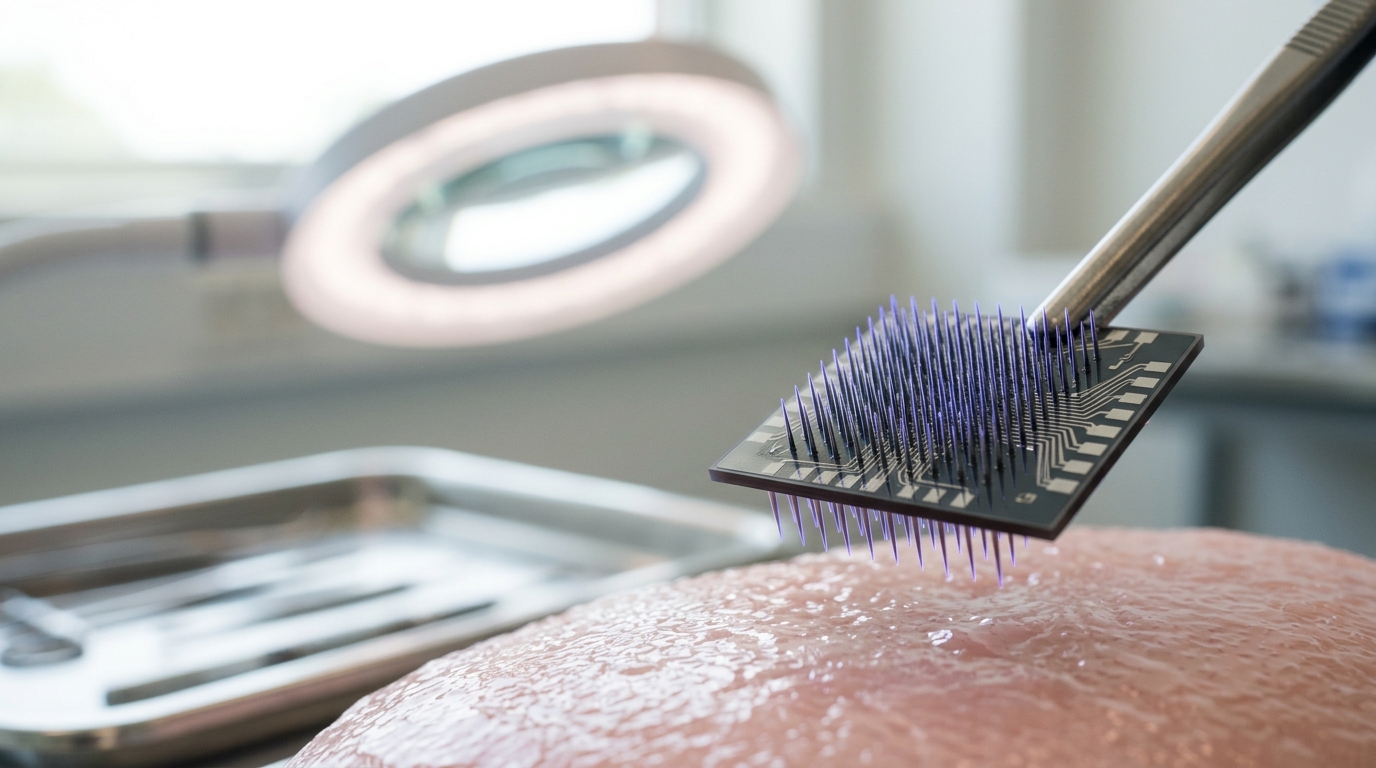
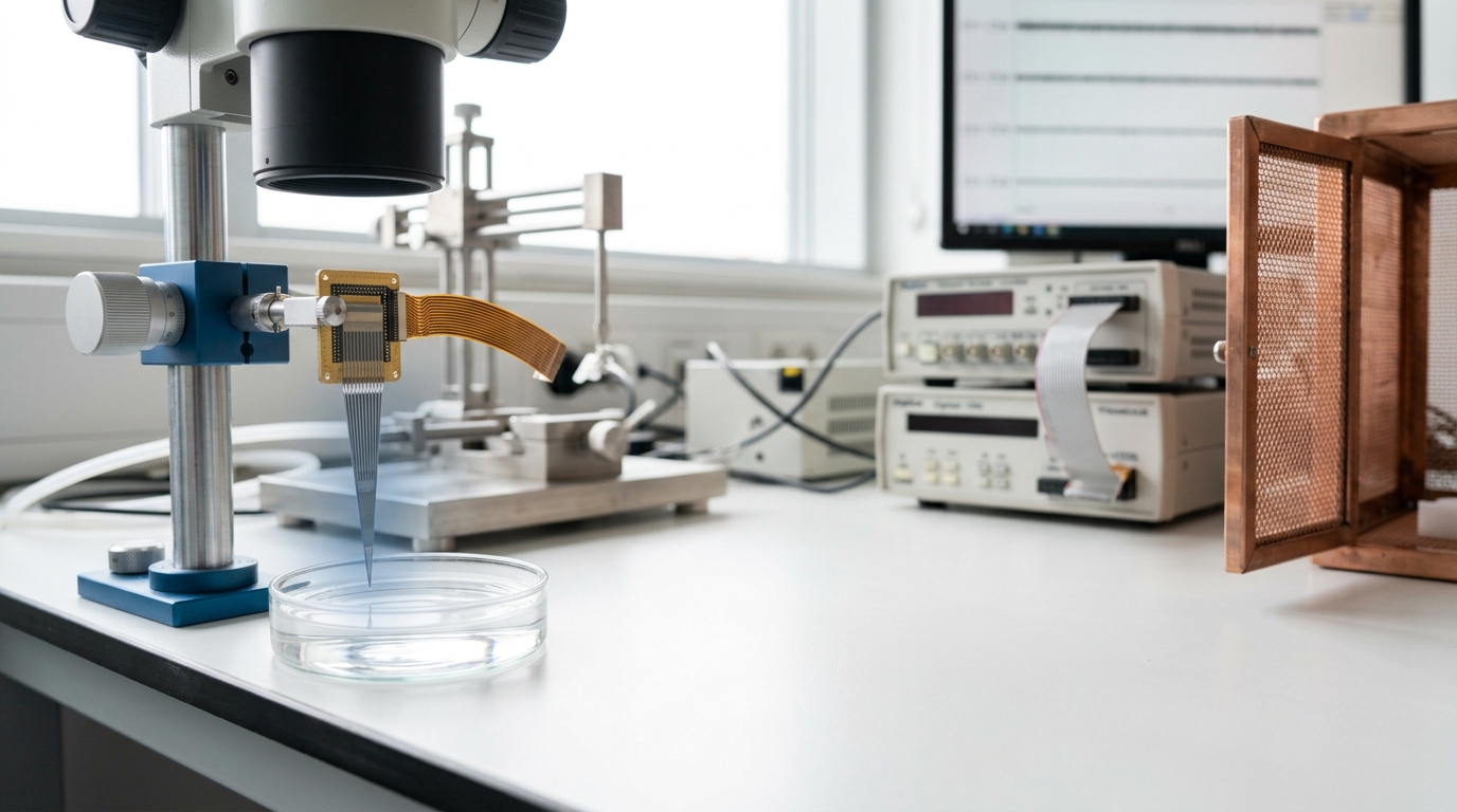
Electrophysiology inserts an electrode, or an array of electrodes, into neural tissue and records the electrical activity of neurons directly — either single-unit action potentials (spikes) from individual identified neurons, or local field potentials reflecting the summed synaptic activity of a surrounding population. The dominant device for high-channel-count recording in both animal research and human clinical brain-computer interfaces is a class of silicon microelectrode arrays, of which the “Utah array” — a rigid grid of roughly 100 needle-like electrode shanks implanted into a few square millimeters of cortex — is the most established. A recent clinical evaluation followed Utah-array recordings across fourteen BrainGate trial participants over periods extending years, characterizing signal stability, electrode yield, and decoding performance over that entire span rather than in a single session [2]. That is the core fact about invasive electrophysiology: temporal resolution at the scale of individual spikes (sub-millisecond), spatial resolution at the scale of individual neurons, but coverage limited to a few hundred to at most a few thousand simultaneously recorded cells, confined to whatever small volume of tissue the array physically occupies, and requiring a surgical implant that in humans is currently justified only by clinical need — paralysis, epilepsy monitoring, or comparable indications — not by scientific curiosity alone.
Non-invasive imaging
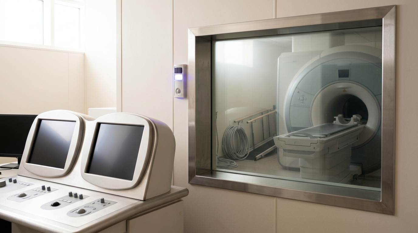
Functional MRI infers neural activity indirectly, from the blood-oxygen-level-dependent (BOLD) signal that follows local changes in blood flow and oxygenation after neural activity increases metabolic demand. Its principal strength is whole-brain coverage at good spatial resolution — millimeters — obtained without surgery, in ordinary healthy volunteers, at scale. The Human Connectome Project’s imaging protocols demonstrate this scale directly: a shared, multimodal acquisition and preprocessing pipeline applied across hundreds to thousands of participants, standardized enough that data collected at different sites can be pooled and jointly analyzed [1]. That population-level reach is something no invasive method can ethically approach in humans. But the cost is real and load-bearing rather than incidental: the BOLD response peaks four to six seconds after the neural event that triggered it, smearing fine temporal structure, and — this is the more consequential limitation — the exact relationship between the hemodynamic signal and the underlying spiking and synaptic activity remains only partially characterized, varies by brain region and cortical layer, and cannot simply be assumed constant across the conditions an experiment wants to compare [3]. Electroencephalography (EEG) and magnetoencephalography (MEG) sit at a different point on the same non-invasive axis: they measure the brain’s electrical and magnetic fields directly, at millisecond resolution, restoring the timing precision fMRI lacks, but at the cost of much coarser spatial localization, because reconstructing where inside the head a scalp- or helmet-level signal originated is a mathematically ill-posed inverse problem with no unique solution [6].
Computational and theoretical modeling
The third family does not record from tissue at all. It builds models — from biophysically detailed single-neuron simulations to abstract population-level dynamical systems to large-scale network models constrained by connectomic data — and tests whether those models reproduce, and ideally predict, patterns actually observed with the other two families. Sejnowski, Koch, and Churchland’s foundational 1988 statement of the field framed this explicitly: the aim is to explain how the electrical and chemical signals available to real neurons can implement the representations and computations that behavior requires, using simulation and theory to bridge levels that no single experiment spans on its own [7]. Modeling’s comparative advantage is unlimited spatial and temporal access — a simulation can report every synapse’s state at every timestep — and the ability to run counterfactual manipulations no living brain permits. Its comparative weakness is equally direct: a model’s output is only as trustworthy as its assumptions, and a simulation that fits existing data can still be structurally wrong in ways that only become visible when it is asked to predict something new.
The three dimensions worth comparing
Spatial and temporal resolution trade against each other, not against a common ideal
This is not a physical law in the way the Heisenberg uncertainty relation is; it is a loose but genuinely useful heuristic for this comparison, worth stating because it exposes the actual trade-off rather than leaving it implicit. No current recording modality achieves fine spatial resolution, fine temporal resolution, and whole-brain coverage simultaneously. Electrophysiology gets sub-millisecond timing and single-neuron spatial precision by giving up coverage — a Utah array samples a few square millimeters [2]. fMRI gets whole-brain millimeter-scale coverage by giving up several seconds of temporal precision to hemodynamic delay [3]. MEG recovers millisecond timing non-invasively but gives up precise localization to the inverse problem [6]. Every real instrument sits on a different point along this trade surface; none sits at the origin.
Invasiveness bounds which questions can be asked of which subjects
Invasiveness is not merely an ethical footnote — it structurally determines the population and therefore the questions a method can answer. Electrophysiology’s need for surgical implantation restricts human use to clinical populations recruited under compelling medical justification, such as the tetraplegic participants in BrainGate trials, whose data are scientifically invaluable but drawn from a narrow, non-representative sample [2]. Optogenetic circuit manipulation — genetically targeting specific neuron types with light-sensitive proteins so a specific circuit element can be switched on or off — is more invasive still, requiring genetic modification, and is accordingly confined to animal models, chiefly rodents, where it has been used to establish which specific circuits causally drive behaviors relevant to anxiety, reward, and fear [4]. Non-invasive imaging inverts this constraint entirely: the Human Connectome Project’s scale — pooling standardized scans across a large, demographically varied healthy population — is possible specifically because MRI and MEG/EEG carry no comparable surgical risk [1].
Causal inference is the dimension most often conflated with the other two
This is the comparison most frequently botched in popular science coverage, so it is worth stating precisely. A method’s causal power is not the same axis as its spatial or temporal resolution, and a method can be excellent on the first two and weak on the third. fMRI, EEG, and passive (non-perturbing) electrophysiological recording are all fundamentally correlational: they observe that a signal changed when a stimulus or behavior occurred, but observing covariation cannot by itself distinguish “this circuit caused the behavior” from “this circuit is a downstream consequence of the behavior” or “both are driven by a common upstream cause” [8]. Optogenetics and, to a lesser degree, focal electrical microstimulation are different in kind, not degree: they intervene on the system and then observe the behavioral consequence, which is the structure an actual causal claim requires. Tye and Deisseroth’s review is explicit that this causal lever — precise enough to target a genetically defined cell type, fast enough to track a specific phase of a behavior — is what optogenetics contributes that no imaging modality can [4]. Jazayeri and Afraz go further and formalize the complication: even a real causal perturbation can be misread if it does not respect the geometry of the circuit’s intrinsic activity patterns, meaning a naive stimulation experiment can produce a result that looks causal without actually isolating the pathway the experimenter intended [8]. Causal power, in other words, is not a simple property a method either has or lacks; it depends on how carefully the intervention is designed relative to what is already known about the circuit’s own dynamics.
Where connectomics and modeling sit relative to both
Connectomics — mapping the anatomical wiring of a brain at the level of individual axonal projections between regions — is neither a recording of activity nor a simulation of it, and it is worth placing separately for that reason. The Allen Institute’s mesoscale connectome of the mouse brain used viral tracing combined with high-throughput serial two-photon tomography to reconstruct axonal projections from defined source regions and cell types across the entire brain, producing an open structural map at a resolution no single electrophysiology or imaging session can match for anatomical completeness [5]. What it does not do is measure activity at all: a wiring diagram tells you what could in principle influence what, not what did influence what on any given trial, and not when. Computational models increasingly use exactly this kind of connectomic structure as a scaffold — constraining a simulated network’s architecture to match measured anatomy before asking whether the resulting dynamics reproduce recorded activity patterns — which is one of the more concrete recent examples of the three families working as a single pipeline rather than three separate literatures.
Fact, overclaim, and open disagreement, kept separate
Fact. Utah-array recordings in BrainGate participants have sustained usable single- and multi-unit signal, with quantified yield and stability, across years of continuous clinical use [2]. The Human Connectome Project’s standardized, multimodal acquisition pipeline has been applied at population scale and its methodological rationale published for scrutiny [1]. The mouse mesoscale connectome is a completed, openly available anatomical dataset [5]. Optogenetic manipulation has established specific causal roles for specific circuit elements in specific rodent behaviors, replicated across multiple laboratories [4].
Vendor-style overclaim, and the shape it usually takes. Popular coverage of brain-computer interfaces and neuroimaging alike regularly elides the difference between “decodes an increasingly rich signal in a controlled clinical or laboratory setting” and “reads thoughts” or “sees what you’re thinking” in any general sense. The clinical BrainGate literature itself is careful to scope its claims to specific decoded outputs — cursor control, typing, in some paradigms speech — under trained, session-specific calibration, not general mind-reading [2]. Similarly, an fMRI finding that a region “lit up” during a task is frequently reported as though it directly demonstrated that region performing a cognitive function, when Logothetis’s methodological critique is precise that the BOLD signal reflects local metabolic and hemodynamic changes whose relationship to spiking output is itself incompletely characterized and region-dependent — a correlate of neural activity, filtered through vasculature, not a direct readout of computation [3].
Genuine, live disagreement. Whether the BOLD signal more closely tracks input synaptic activity (local field potentials) or output spiking in a given brain region and cortical layer remains an active empirical question rather than a settled fact, and Logothetis’s review is explicit that this is a limitation of what is currently known about neurovascular coupling, not a problem solvable simply by building a more powerful scanner [3]. Separately, how to correctly attribute a causal role from a perturbation experiment — when a stimulation-evoked change in behavior reflects the targeted circuit’s normal function versus an artifact of activity patterns the intervention itself imposed — is treated by Jazayeri and Afraz as an open methodological problem requiring a formal framework, not a solved question with an agreed procedure [8].

Convergence, and a bounded prediction
The clearest current trend is not any one method displacing the others but three families converging on the same experiments: simultaneous electrophysiology and fMRI in the same animal to directly calibrate what the BOLD signal reflects at the neural level, connectome-constrained computational models tested against activity recorded from the regions they simulate, and closed-loop brain-computer interfaces where a decoded model of neural activity is used, in real time, to select the next stimulation. None of these convergent designs make any single method’s core trade-off disappear — a multimodal experiment still pays fMRI’s temporal cost during the imaging component and electrophysiology’s coverage cost during the recording component; it just runs both at once on the same tissue.
Prediction, with a horizon and a disconfirmation condition. Over the next five to ten years, expect the primary contribution of large-scale connectomic and multimodal-recording datasets to be sharper constraints on which circuit-level models are viable, rather than a wholesale replacement of single-method studies with combined ones — because the surgical, financial, and regulatory cost of simultaneous multimodal recording in humans will keep it rare relative to single-modality studies for that whole window. The observable indicator to watch is the ratio of published studies using genuinely simultaneous multimodal recording (not merely the same subjects at different sessions) to single-modality studies in high-impact systems neuroscience venues. If that ratio has not measurably risen by the early 2030s relative to its current level, the prediction that convergence is the field’s leading edge — as opposed to a promising but still-marginal niche — should be treated as disconfirmed.
What “comparing approaches” should not mean
None of the foregoing supports ranking electrophysiology, imaging, and modeling against one another on a single scale, and the temptation to do so should be resisted specifically because the three dimensions compared here — resolution, invasiveness, and causal power — do not collapse into one number. A method that is unmatched on temporal and spatial precision but confined to a few hundred neurons in a clinical population answers a different question than a method that is unmatched on population coverage but blind to individual spikes, and both differ again from a method that has no access to real tissue at all but can test a hypothesis no living experiment can run. Systems and computational neuroscience makes progress by matching the method to the question, not by waiting for one instrument to subsume the other two.